Te Papa / October 2008
November 2008 | September 2008-
-

Mounting a squid
- Te Papa's blog
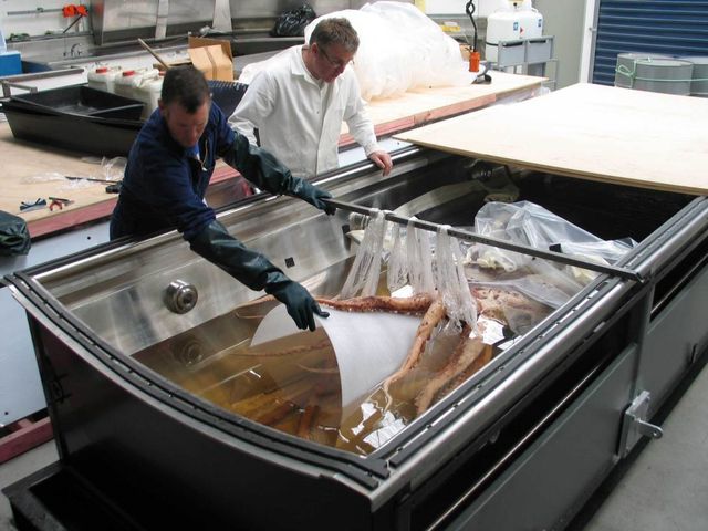
- Work is underway preparing the mounting system for the colossal squid in its display tank. Mark kent and Robert Clendon preparing the squid for exhibition. Image copyright Te Papa Unless the squid is supported by acrylic mounts it will remain a collapsed heap on the bottom of the tank - not very appealling! To display it in as realistic pose as possible a series of supports will be manufactured to splay the tentacles out so the beak can be seen, and to expand the mantle from its collapsed position. The squid will be slightly angled to one side in the tank, so it will be possible to see the eye and even the funnel, which is on the under side of the mantle. lighting inside the tank will illuminate the specimen from the sides - eliminating any glare or reflection from the surface of the preservative. Te Papa conservators Mark Kent and Robert Clendon have to work with the specimen partially supported by the liquid preservative. The arms are held in the desired position using plastic food wrap while they prepare the template. The template will then be used to cut the final acrylic mount. Once the templates are completed and the mounts made, the specimen will be moved to the museum building at Cable Street and mounted properly, before the lid is placed on the tank it is filled with the preservative solution. Preparing the mount template. Image copyright Te Papa Museum conservators and mount makers have to deal with objects ranging from artefacts to fine art sculptures. The colossal squid is a new challenge!       
- Automatically tagged as:
- blogs
- te-papa
-
-
-

Rare fern rediscovered.
- Te Papa's blog
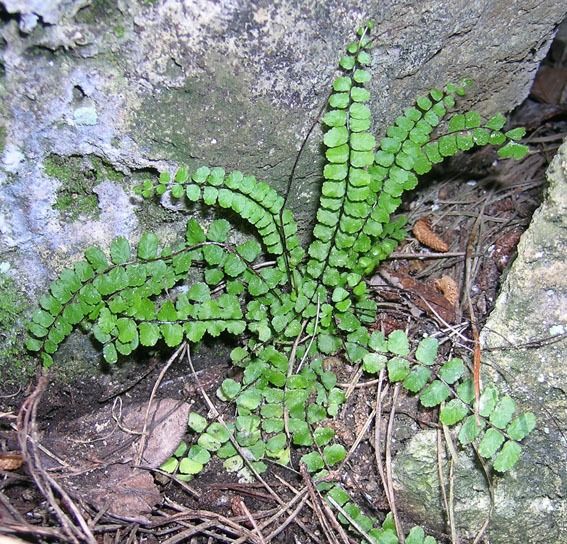
- I’m one of the Botany Curators at Te Papa, and ferns are one of my specialties. New Zealand has about 200 native ferns, and some of them are very rare. We recently rediscovered one rare fern that had been ‘lost’. I was beginning to wonder if it had become extinct, but fortunately it has not. Still, the known total of individuals is still only 9, and this population is only a goat-lunch away from extinction! Me, on top of the Ruahine Ranges. No rare ferns sighted up here, but interesting nonetheless. The rediscovered fern is a maidenhair spleenwort. It had been definitively identified from just three New Zealand sites, all in Hawke’s Bay, and all dating to the 1950’s. The localities of these three sites were not precisely recorded, and no one I talked to knew of a living population. I enlisted the help of the Manawatu Botanical Society to search one of these sites (the most precise one, which involved searching several square km rather than several tens of square km). I wasn’t very optimistic, given the amount of time since it had been previously collected and that I had already looked at a number of similar Hawke’s Bay sites. But, we found it - 9 plants in one very small area. There is another maidenhair spleenwort in NZ, and it is quite common. These two maidenhair spleenworts look similar, but they have different chromosome numbers; the common one has six sets of chromosomes while the rare one has four sets. This kind of difference is usually treated at the subspecies or even species level in ferns. Unfortunately, the present taxonomy, or formal scientific naming, for these ferns is not adequate. We hope to sort this out in the next year or so. They have both been called Asplenium trichomanes, but this fern does not occur in NZ (at least when interpreted in a narrow sense). The rare maidenhair spleenwort in NZ has also been called Asplenium trichomanes subsp. quadrivalens; whether this is correct remains to be established. Maidenhair spleenwort. This is the rare species, but the common one looks very similar. The two maidenhair spleenworts usually occur on or near limestone. They can be distinguished from all other ferns in NZ by their undivided, black, almost smooth stems, and by having their reproductive structures in lines away from the margins of the undersides of their leaves. This particular arrangement of the reproductive structures characteristics all of the spleenwort (Asplenium) species, of which there are about 20 in NZ (and some 600 in the world). I’d be interested in learning of additional maidenhair spleenwort sites in Hawke’s Bay. Both species of maidenhair spleenwort have been recorded from the Hawke’s Bay, so any new finds may be the rare or the common species. I would need to closely inspect them to be sure. But, please, do not remove them from the wild! Email (leonp@tepapa.govt.nz) or phone (04 381 7261) me the locality details. Te Papa’s Collections Online includes a photo of a maidenhair spleenwort specimen collected from the Hawke’s Bay in 1881 (it’s the common species, rather than the rare one). The New Zealand Plant Conservation Network also has more information about maidenhair spleenworts.       
- Automatically tagged as:
- blogs
- te-papa
-
-
-

Squid - the inside story
- Te Papa's blog
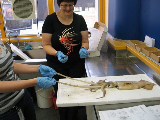
- It’s a lovely spring Friday morning in Wellington. What else would we (Pamela, Chris and Judy - our brave and newest squid team member) be doing other than dissecting a couple of nice fresh squid from the local wholesale fish supplier? It’s all in the interest of bringing you a bigger and better exhibition on the colossal squid, as we come to grips with squid anatomy (literally). We quickly discovered that not all squid are the same on the inside (surprise) and that once inside them it can be a messy business. Note to self - try to avoid puncturing the ink sac until the end. We started with an arrowsquid. We checked the arms and the tentacles - all eight arms and two tentacles present and correct. The suckers on the arms had hard little circles, which pop out - who needs to pop bubble wrap? Cutting through the mantle was hard work - you need a sharp pair of scissors or a good scalpel. Pulling back the folds of the mantle reveals the inner organs. Working out what they all are is a challenge but we think we identified the gills, the stomach and caecu,m and what we thought was the hearts. Yep, that’s right, a squid has three hearts. The eyes were exciting to dissect. It was a thrill to extract the lenses and find that they come in two parts - just like the colossal squids, and indeed all squid. The arrowsquid lens is a lot smaller - around 0.5cm across - compared with the huge orange-sized lens of the colossal. It was also really exciting to remove the beakfrom the really dense muscular tissue surrounding it. First we got the lower beak out, then the upper beak and we could see how they fit together. Then we came across the radula - it’s a bit like a tongue - with it’s amazing rows of sharp, raspy teeth. Stomach contents of our squid were examined. We could feel the crunchy bits inside, and these turned out to be fish vertebrae. Last but not least we cleaned away all the messy bits to expose the mantle - and extract the gladius, or pen. This incredible structure just glides out of the mantle and looks for all the world like it’s made of plastic. So I’m hooked on squid anatomy - there will be photos and more on the broad squid we examined next, shortly.       
- Automatically tagged as:
- blogs
- te-papa
-